
Tumor Staging
Staging helps doctors determine how serious the cancer is and how best to treat it. It describes the severity of a person’s cancer based on the magnitude of the primary tumor as well as the extent the cancer has spread to other areas of the body. Staging is generally based on the guidelines from the American Joint Committee on Cancer -TNM staging system. You are likely to see your cancer described by this staging system in your pathology report.
The TNM is based on the size and/or extent of the primary tumor (T) and how far it has spread within the body, where the malignancy has moved to the regional lymph nodes (N), or distant sites within the body (M) such as the lungs, liver or kidney.
When your cancer is described by the TNM system, there will be numbers after each letter that give more details about the cancer. Based on these three elements, a tumor is then classified as being Stage 1, 2, 3 or 4 with stage 4 being the most serious. Many ACC patients are classified as stage 4 but due to the slow growth of this cancer, patients are living longer lives vs other common cancers.
For example, a tumor classified as T3NOMO refers to a large tumor that has not spread to the lymph nodes (NO) or any other part of the body (M0). The following explains what the letters and numbers mean:
Primary tumor (T)
TX: Main tumor cannot be measured.
T0: Main tumor cannot be found.
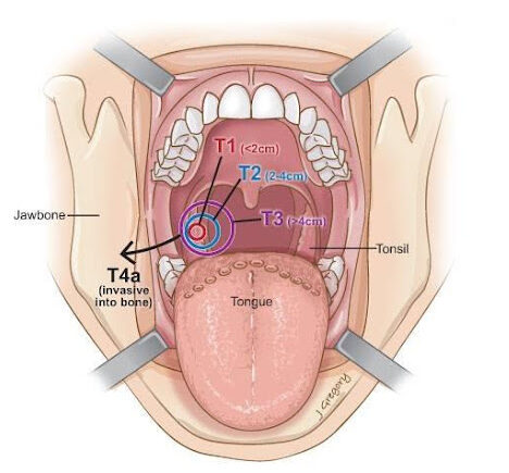
T1, T2, T3, T4: Refers to the size and/or extent of the main tumor. The higher the number after the T, the larger the tumor or the more it has grown into nearby tissues.
Distant metastasis (M)
M0: Cancer has not spread to other parts of the body.
M1: Cancer has spread to other parts of the body.
The TNM system helps describe cancer in great detail but doctors may look at other information about ACC for clues about how it will behave
Regional Lymph Nodes (N)
N0: There is no cancer in nearby lymph nodes.
N1, N2, N3: Refers to the number and location of lymph nodes that contain cancer. The higher the number after the N, the more lymph nodes that contain cancer.
Tumor Grade
When examined under a microscope, ACC tumors are characterized by a distinctive histological pattern of abnormal “nests” or cords of certain cells (epithelial cells) that surround and/or infiltrate ducts or glandular structures within the affected organ. These structures are typically filled with a mucous-like material or contain abnormal fibrous membranes. ACC is almost always considered a low-grade malignancy that has a history of slow growth.
In a smaller number of cases, high grade adenoid cystic carcinoma means that the tumor may consist (or change) in a way that results in a more aggressive behavior. ACC tumors consist of 3 cell patterns or types when examined under a microscope – cribriform, tubular and solid. A ‘solid’’ histology or 30% or more of this type may indicate a high grade ACC transformation. High grade transformation is important because these tumors may be more likely to metastasize (spread) to the lymph nodes and other areas of the body particularly the lungs.